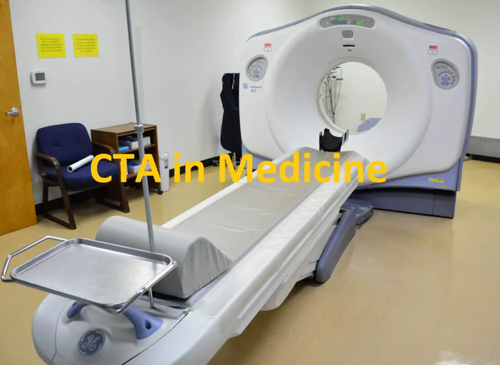
CTA in medical terms/ abbreviation stands for computed tomography angiography, which is a medical imaging technique that uses computed tomography (CT) to visualize the blood vessels and tissues in different parts of the body
How CT Angiography works
CT is a type of X-ray that uses a computer to create cross-sectional images of the body. By injecting a special dye called contrast material into the bloodstream, CT can enhance the visibility of the blood vessels and tissues. The contrast material makes the blood vessels appear brighter on the CT images, allowing the radiologist to see their shape, size, and condition
CTA was first introduced in the 1980s, but it became more widely used in the 1990s with the development of spiral or helical CT scanners. These scanners can acquire images faster and with higher resolution than conventional CT scanners, making CTA more accurate and reliable.

Types of CTA and uses
CTA can be used to examine blood vessels in many key areas of the body, such as the brain, heart, lungs, kidneys, abdomen, pelvis, and limbs. Depending on the area of interest, different types of CTA can be performed. Some examples are:
- Coronary CTA: This type of CTA is used to assess the arteries of the heart and detect any blockages, narrowing, or abnormalities that may cause chest pain or increase the risk of a heart attack
- Aortic CTA: This type –of CTA is used to evaluate the aorta and its major branches, such as the carotid, renal, and mesenteric arteries. It can detect any aneurysms (ballooning), dissections (tearing), or stenosis (narrowing) that may affect the blood flow or cause complications
- Pulmonary CTA: This type of CTA is used to examine the lungs and the pulmonary arteries that supply them with blood. It can diagnose pulmonary embolism (blood clots in the lungs), pulmonary hypertension (high blood pressure in the lungs), or other lung diseases
- Peripheral CTA: This type of CTA is used to study the blood vessels in the arms and legs. It can identify any blockages, narrowing, or damage that may cause pain, swelling, or poor circulation. It can also help plan for vascular surgery or interventions
Pros and Cons of CTA
CTA has many advantages over other imaging modalities, such as ultrasound, magnetic resonance angiography (MRA), or conventional catheter angiography. Some of these advantages are:
- CTA is fast and noninvasive. It can be done in minutes without requiring any anesthesia or sedation. It does not involve inserting any catheters or wires into the blood vessels
- CTA is accurate and detailed. It can produce high-resolution images that show both the blood vessels and the surrounding tissues in three dimensions. It can also measure the diameter, length, and angle of the blood vessels
- CTA is widely available and cost-effective. It can be performed in most hospitals and imaging centers using standard CT scanners. It does not require expensive equipment or specialized staff. It is also cheaper than catheter angiography
However, CTA also has some limitations and risks that need to be considered. Some of these are:
- CTA exposes the patient to radiation. Although the amount of radiation used in CTA is minimal and comparable to other X-ray procedures, repeated exposure may increase the risk of cancer in some patients. Therefore, CTA should be done only when necessary and with appropriate precautions
- CTA requires contrast material injection. Some patients may have an allergic reaction to the contrast material or experience side effects such as nausea, vomiting, or itching. The contrast material may also damage the kidneys in some patients with kidney disease or diabetes. Therefore, patients should inform their doctors about any allergies or medical conditions before undergoing CTA
- CTA may not be suitable for some patients or situations. For example, patients with metal implants, pacemakers, or other devices may not be able to have CTA because they may interfere with the CT scanner. Patients who are pregnant, obese, claustrophobic, or unable to hold their breath may also have difficulties with CTA. In some cases, other imaging modalities may provide better results than CTA. Therefore, patients should discuss with their doctors about the best option for their specific needs
Difference between Normal CT and CTA.
Conventional Computed Tomography (CT) and CT Angiography (CTA) are both valuable medical imaging techniques that utilize X-rays to create detailed cross-sectional images of the body. However, they have distinct differences in terms of their applications, processes, and the information they provide.
1. Purpose and Applications:
- Conventional CT: Conventional CT scans are primarily used to visualize and diagnose a wide range of conditions within the body, such as detecting tumors, assessing injuries, and identifying abnormalities in organs or bones. It provides detailed images of anatomical structures and can help doctors plan surgeries or monitor the progression of diseases.
- CT Angiography (CTA): CT angiography, on the other hand, is specifically designed to visualize blood vessels and the flow of blood within them. It is often used to diagnose conditions affecting the blood vessels, such as aneurysms, stenosis (narrowing of blood vessels), and vascular malformations. CTA can provide crucial information about blood vessel health and identify potential blockages or abnormalities.
2. Contrast Enhancement:
- Conventional CT: In a conventional CT scan, a contrast agent may be injected into the patient’s bloodstream to enhance the visibility of certain structures or abnormalities. This helps differentiate various tissues and organs, making them stand out more clearly in the images.
- CT Angiography: CTA heavily relies on the use of contrast agents to visualize blood vessels. The contrast dye is injected intravenously, and as it travels through the blood vessels, it allows the radiologist to capture detailed images of the vasculature and detect any issues like blockages or aneurysms.
3. Imaging Process:
- Conventional CT: In a conventional CT scan, a rotating X-ray machine captures cross-sectional images as it moves around the patient’s body. The data is then processed by a computer to create detailed images of the scanned area.
- CT Angiography: CTA involves a similar process of capturing cross-sectional images, but it focuses on the blood vessels. The contrast dye’s movement through the vessels is tracked in real-time, allowing the creation of dynamic images that show both the anatomical structures and the flow of blood.
4. Radiation Exposure:
- Conventional CT: Both conventional CT and CTA involve exposure to ionizing radiation, which can be a concern, especially when multiple scans are necessary. Radiation exposure can vary depending on the type of scan, the area being imaged, and the individual’s specific circumstances.
- CT Angiography: CTA typically requires a higher dose of contrast and, consequently, a higher dose of radiation compared to conventional CT. The benefits of the information obtained through CTA usually outweigh the radiation risks, especially in cases where vascular conditions are suspected.
In summary, while both conventional CT and CT Angiography utilize X-rays and computer processing to create detailed images, their primary purposes and applications differ significantly. Conventional CT scans focus on imaging various body structures for diagnostic purposes, whereas CT Angiography specializes in visualizing blood vessels to diagnose vascular conditions. The use of contrast agents and radiation exposure levels are also notable differences between the two techniques. The choice between conventional CT and CTA depends on the specific medical condition being investigated and the information needed for accurate diagnosis and treatment planning.
Reference:
Rankin SC. CT angiography. Eur Radiol. 1999;9(2):297-310. doi: 10.1007/s003300050671. PMID: 10101654.
